Ch1 Fundamentals
Copyright © 2025 Mark Song
Homeostasis: Hold at a steady state.
Negative feedback mechanisms are crucial in regulating homeostasis
Feedback Loops:
Negative Feedback: (more common)
The system is composed of four parts, sensor, controlled variable, effector, and controller.
The controller has a set point and the controller has a comparator which compares with the sensor input and it will send an error signal (within the controller) and actuating signal (exits the controller) to effectors.
e.g., Keep warm in winter:
sensor is the thermometer, the controlled variable is the temperature, the controller is the thermostat, the effector is the furnace/heater
if the room temperature are no smaller than the set point, the error signal is 0/+ and the actuating signal will not be sent
e.g., body temperature, mood, plasma concentration of (H2O, H+, Ca2+, Glucose)
e.g., body temperature
controlled variable is the body temperature
sensor is the thermoreceptors
controller is the hypothalamus
effectors are the skeletal muscles, skin arterioles (thin blood vessel on skin, which dilation would cool down body temperature), sweat glands, etc..
Positive Feedback:
The system is composed of four parts, sensor, output variable, effector, and amplifier.
no controlled variable or set point
properties:
plateau, maximum value of output of effector
threshold, level of output variable at which point system rapidly drives towards plateau
e.g., Action potential (V-gated Na channels),
e.g., labor and childbirth
output variable: pressure in cervis and uterine wall
sensor: pressure sensitive sensory neurons in cervix and uterine wall
amplifier: hypothalamus/pituitary
actuating signal: oxytocin
effector: uterine smooth muscle
plateau: the maximum force that the uterine smooth muscle can generate
Transport
three primarily focused factors that drives movements
pressure (heart pumping blood), concentration (diffusion), electrical charge (v-gated channels)
transport across semipermeable membrane
w/o protein: oxygen gas, steroid hormones, water (in small amount)
w/ protein: ions, glucose, water (in large amount)
Water diffusion aka osmosis, isotonic is ok, hypotonic is more solute in cell and the cell will swollen, hypertonic is less solute in the cell and the cell will shrink
factors for ion transport
concentration gradient
charge gradient
For a real cell:
K concentration is higher inside the cell and Na concentration is lower inside the cell
K is more closer to equilibrium
Ways of transports:
- Passive
- Simple Diffusion
- Diffusion via channel
Channel allow diffusion through membrane
- ion channels
- aquaporins
- uniporter carrier protein
allow passive movement
- Active
- Primary active transport
e.g., Na/K ATPase (3Na out, 2K in) took 30% of all energy
- Secondary active transport
e.g., SGLT1 and SGLT2 are Na/glucose co-transporter (Na down the gradient, glucose up the gradient)
- Primary active transport
- endocytosis
engulfing extracellular substances
internalization of transmembrane proteins
- exocytosis
Secretion of proteins or other messengers
trafficking of transmembrane proteins to membrane
Cell-to-cell communication
The chemical messenger/ligand/first messenger binds to a specific receptor molecule
Ligand type:
- Lipid soluble messager — binds to receptors in cytoplasm or nucleus
- Water soluble messager — binds to the receptors on the cell membrane. e.g., ionotropic receptors (nAchR)
GCPRs G-Protein Coupled Receptors
- Receptor associated with heterotrimeric G protein (alpha, beta, gamma)
- Ligand binding causes alpha subunit to bind GTP instead of GDP
- GTP binding causes the alpha subunit to dissociate from beta, gamma subunit
- The alpha subunit interacts with and activates a transmemebrane enzyme or ion channel
- The alpha subunit has intrinsic GTPase activity, turning itself off by catalyzing cleavage of the third P in GTP
- Subunits reassoicate with each other and receptor
e.g., oxytocin binds with GPRC
e.g.
- G alpha subunit activates adenylyl cyclase
- Adenylyl cyclase catalyzes production of cAMP from ATP
- cAMP binding activates cAMP- dependent protein kinase (also called Protein Kinase A (PKA))
Key notes:
- Importance of second messengers in signaling pathways
- Amplification of a signal
- Key role of kinases in many signaling pathways
Hormone: a secreted molecule that travels through the blood to target cell(s), exerts some effect based on interaction with receptor
Some hormones can acts as NeruoTransmitters
Endocrine System
Are one of the two main physiological control system. (with nervous system)
Roles in widely varying physiological function such as homeostatic regulation of ions, energy availability, and coordinated changes such as growth and development.
Comparisons between Endocrine and Nervous System
NS are targeted at one cell via synapse, fast, shorter duration
ES are by the hormone that are secreted by endocrine gland, more cells, slower, longer duration
Hypothalamus (with pituitary gland below it)
Hypothalamus have neurosecretory cells whose axons release hormones into capillaries in posterior pituitary, and neurosecretory cells whose axons release hormones into portal system that transports blood a short distance to anterior pituitary.
A Stress Response pathway
Stress (such as psychosocial stress, temperature, fasting, exercise, and anything that are a threats to homeostasis)
hypothalamus release Corticotropin Releasing Hormone (CRH)
anterior pituitary release AdrenoCorticoTropic Hormone (ACTH)
adrenal cortex release cortisol
most tissues response to cortisol
Adrenal gland releases several hormones
Inner most section: Medulla
Releases epinephrine and norepinephrine
Outer section: cortex
Inner: Zona Reticularis
releases androgens and small amount of cortisol
Mid: Zona fasciculata
releases cortisol and small amount of androgens
Outer: Zona glomerulosa
release aldosterone
CRH, ACTH are water soluble and Cortisol are lipid soluble
Cortisol
- Stimulation of liver cell uptake of amino acids and conversion to glucose.
- Stimulation of triglyceride breakdown in adipocytes.
- Inhibition of inflammation.
- Inhibition of nonessential functions (e.g. growth and reproduction).
Inhibits CRH and ACTH release and thus inhibit cortisol release
Insulin Pathway
Sensor: Beta islet cells in pancreas
Controller: Beta islet cells in pancreas
Actuating signal: Insulin
Effectors: Skeletal muscle, adipocytes, liver cells
Controlled Variable: blood plasma glucose level
Beta islet cells has Glut2 transmembrane protein (glucose transporter protein)
Glucose would be converted into ATP
ATP binds to ATP-sensitive K+ channel ()
K+ stop flowing out of the channel
This cause an increase in cell potential
This cause V-gated Ca2+ channels to open
Ca2+ flows into the cell (positive feedback loop in increasing cell potential)
Exocytosis release insulin
Insulin would activate insulin receptor (transmembrane, tyrosine kinase), which would stimulate Glut4 exocytosis and thus increase Glut4 intake. (skeletal muscle)
Insulin would activate insulin receptor, which initiate production of glycogen (from glucose, glycogenesis, which glucose if transported into the cell via Glut2) (liver cell)
Glucagon pathway
Sensor: Alpha islet cells in pancreas
Controller: Alpha islet cells in pancreas
Actuating signal: Glucagon
Effectors: Liver cells
Controlled Variable: blood plasma glucose level
Glucagon binds to glucagon receptor (GPCR), increase cAMP, produce glucose from glycogen (glycogenolysis), glucose flows out of the cell via Glut2.
Diabetes Mellitus
Plasma glucose level are very high
Type 1, problem with beta islet cells, that not enough of insulin produced (might be because of auto-immune diseases)
Type 2, insulin insensitivity in skeletal muscle, adipocytes, and or liver cells
Ca2+ is important
- Insulin release
- Parathyroid hormone signaling
- Neuronal signaling
- Muscle contraction
Ca2+ regulations
includes
- Parathyroid glands
- Bones
- Kidneys
- GI tract
Negative feedback loop
Controlled variable: plasma levels of Ca2+
Sensor: Parathyroid gland cells via CaSR (Calcium Sensing Receptor)
Controller: Parathyroid gland
Actuating signal: PTH
Effectors:
- Bone
Osteoclast break down calcified extracellular matrix in bone tissue, which releases Ca2+
- Kidney epithelial cells
increase Ca2+ reabsorption
- Kidney endocrine cells
release 1,25 dihydroxy vitamin D (lipid soluble), binds to Vitamin D Receptor (VDR) (in intestine) and it enter the nucleus, and turns up TRPV6 transcription, and TRPV6 would merge with the cell membrane and it is a Ca2+ channel that intakes Ca2+
Parathyroid glands:
Located near the throat
Express a transmembrane receptor called Calcium Sensing Receptor (CaSR)
CaSR is GPCR
Ca2+ binds to CaSR and initiates a pathway that inhibit ParaThyroid Hormone (PTH) release
PTH is hydrophilic hormone that binds to GPCR
PTH pathway
- Adenylyl Cyclase / cAMP / ProteinKinaseA pathway
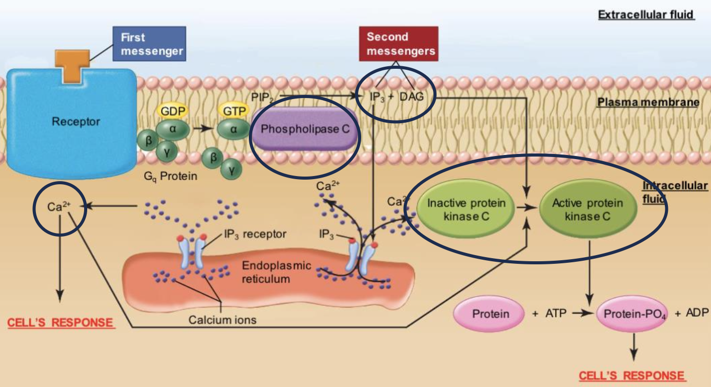
- Phospholipase C Pathway
- Activates Phospholipase C
- Activate IP3 (release Ca2+ from ER and ), DAG (activate Protein Kinease C), Ca2+ (released by Ca2+)