Ch4 Cardiovascular System
Copyright © 2025 Mark Song
Blood vessles
Arteries away from heart
veins to the heart
Pulmonary
right ventricle to left atrium
Systemic
Left ventricle via aorta to right atrium via vena cava
Right hearted are less oxygenated
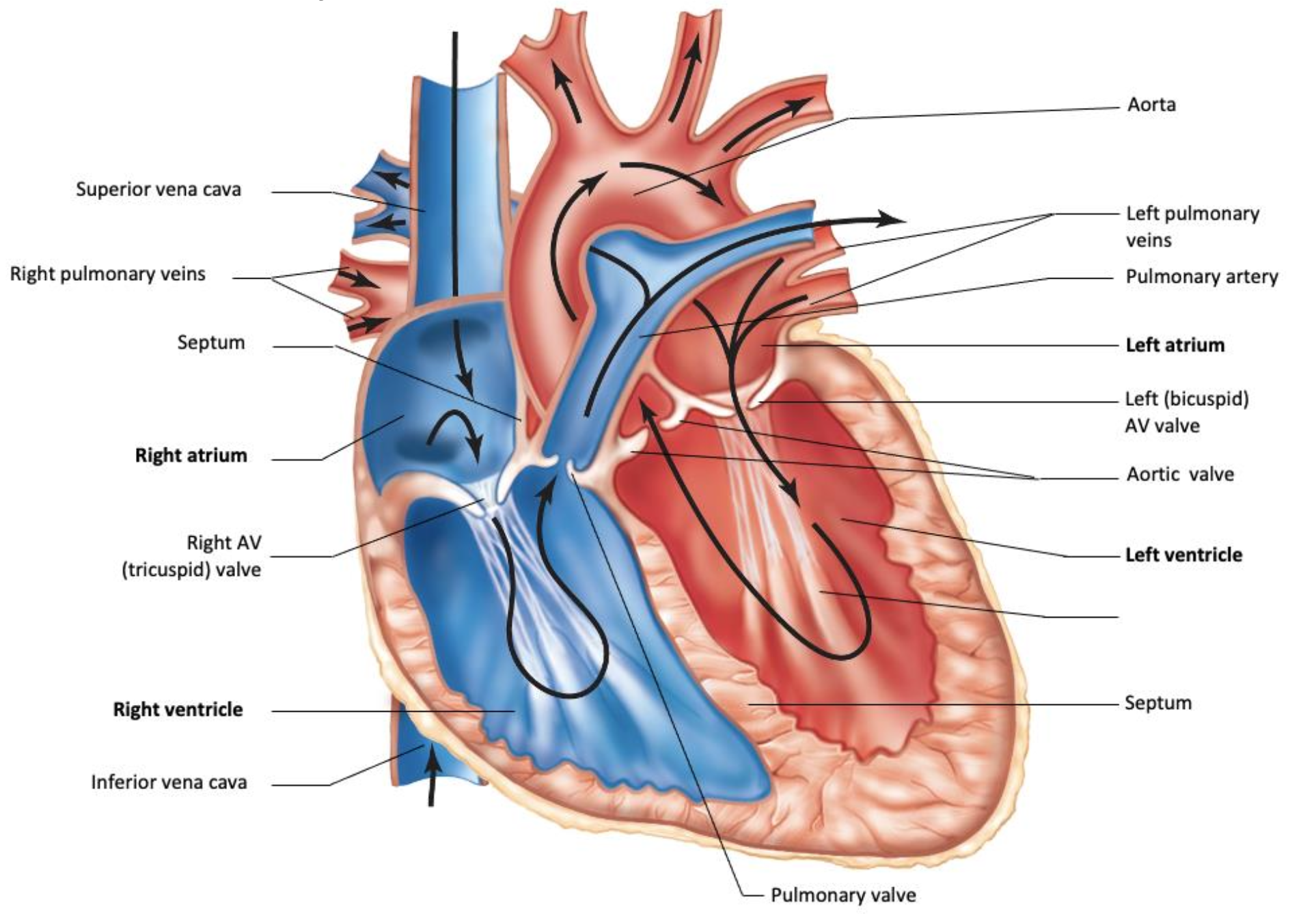
Blood move is moved via bulk flow
Process of blood
- blood enters atria from veins
- AV values open
- blood enter ventricle (80% full of blood)
- atria contract (remaining 20%)
- atria relax, ventricle contracts, AV value closes (lub)
- Pulmonary / aortic value opens
- Pulmonary / aortic value closes (dub)
Electrical signaling
Cells:
contractile cells
Contracts when receive signal
Conducting cells
Does not contracts
Controlled the the Cardiac Conduction System (CCS)
CCS can generate and pump on its own
SinoAtrial (SA) node is the node on the right atrium
then go through inter-nodal pathway
then go to AtrioVentricular (AV) node on the right atrium which delay for 100ms
then go through bundle of his
then go to left and right ventricle and spread out via Purkinje fibers
P wave voltage increase in atrial muscle cell
QRS wave ventricular contraction
T wave ventricular relaxation
Hyperpolarization-activated Cyclic Nycleotide-gated channel 4 (funny channel, HCN4)
HCN4 opens when voltage decrease
cause autorhythmicity
opens when V is below -40mV and with cAMP
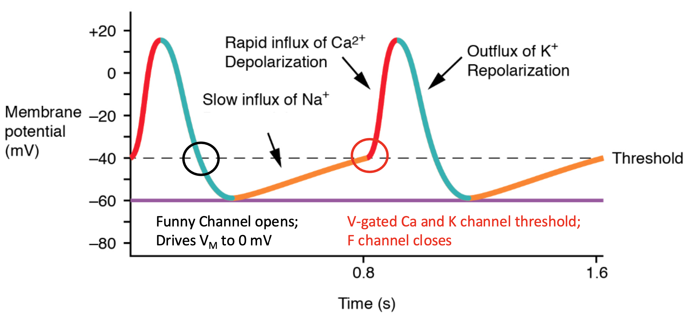
pacemaker potential is the slow influx of Na
Cardiac muscle cells does not have leaking Na channels
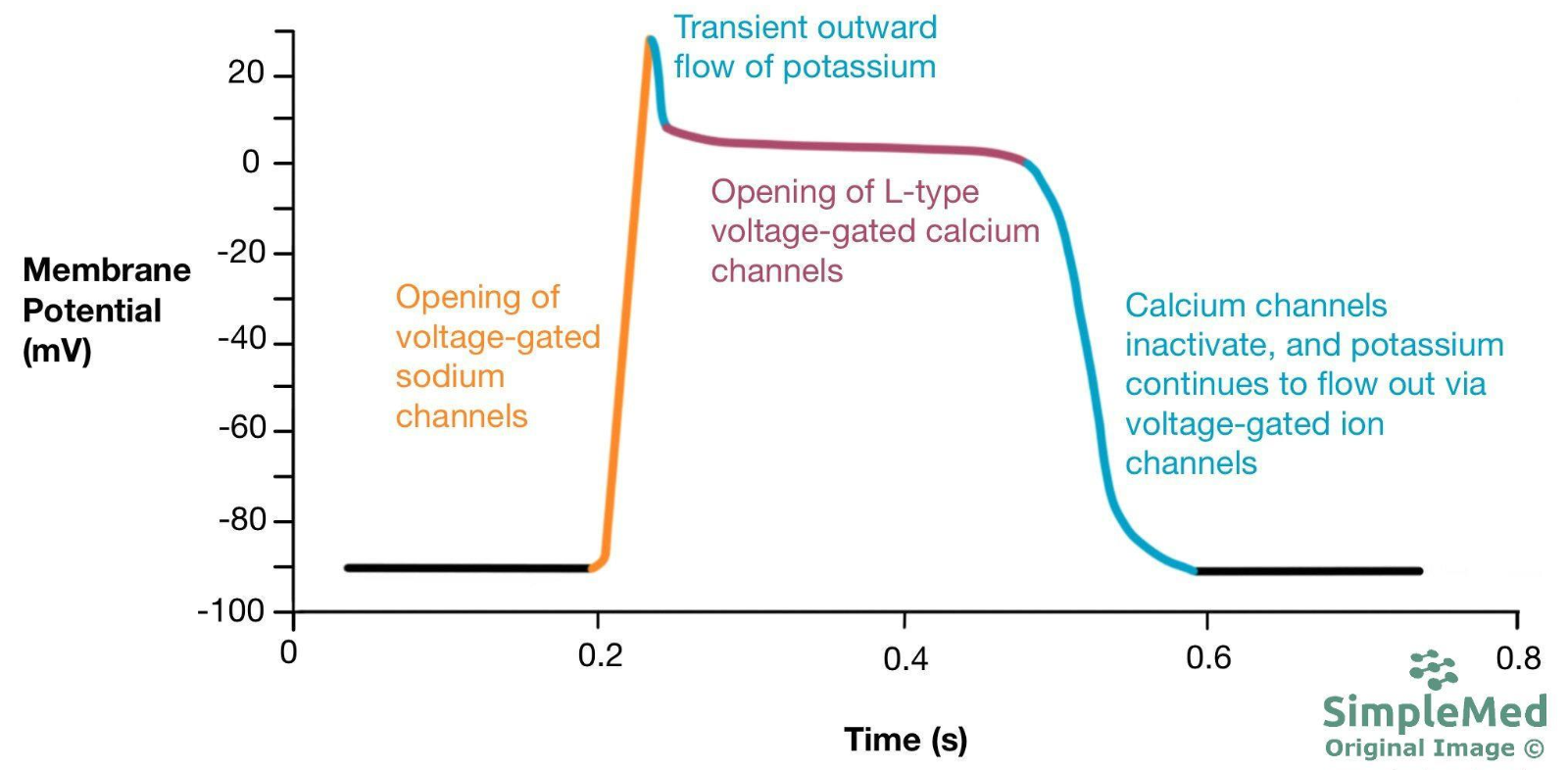
The upward is sodium, which is the start of P wave (atria) or QRS (ventricle)
The first downward is K, which the end of P wave (atria) or QRS (ventricle)
The plateau is the gap between P and QRS or QRS and T
The last downward is K, which is QRS (atria) or T (ventricle)
Layers of Large Artery
Lumen
Endothelium
multiple elastic layer
many layer of connective tissue and muscle
Arteriole
Lumen
Endothelium
many layer of muscle
Capillary
Lumen
Endothelium, with intracellular clefts with proteins unable to flow through
Venule
Lumen
Endothelium
Large vein
Lumen (wide)
Endothelium
Few elastic layers
Few smooth muscle and connective tissue
with valves
Mean Arterial Pressure (MAP) = Cardiac output (CO) times Total Peripheral Resistance (TPR)
MAP feedback loop
Controller: Medulla Oblongata
Sensor: Mechanoreceptor neurons
Actuating signal:
Parasympathetic firing rate
Sympathetic firing rate
Effector
SA node (beta adrenergic for SNS, nAchRs for PNS)
Arteriolar smooth muscle (alpha adrenergic)
SNS: Noradrenaline
| SA node | Arteriole Smooth Muscle | |
| Receptor | beta adrenergic receptor (GPCR) | alpha adrenergic receptor (GPCR) |
| Leads to | Increase cAMP on F channel increase heart rate and CO | Increase smooth muscle contraction Increase TPR |
PNS: Ach
| Heart | |
| Receptor | Muscarinic Ach Receptor, mAchR |
| Leads to | Drop cAMP Increase K conductance decrease heart rate and CO |
Local change
When there is high level of cellular activity, it leads to more blood flow of that area (active hyperemia)
increase in arteriole diameter increase