Ch5 Respiratory system
Copyright © 2025 Mark Song
Trachea, then bronchus, bronchioles, terminal bronchioles, respiratory bronchioles, alveolar ducts, alveolar sacs
The lung’s bottom is attached to the diaphragm
The lung lay in the chest (plural) cavity, which is separated into two
The lung has visceral pleura, intraplerual fluid, and parietal plural outside of it
Inspiration, air in
Expiration, air out
Driven by diaphragm (contract) and external intercostal muscle
alveoli, lung capillaries, tissue capillaries, cells
The binding curve of Hemoglobin
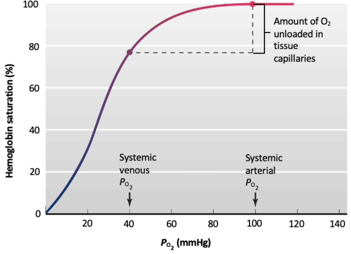
There are four subunits of Hb, and binding of 1 unit of O2 makes binding of more O2 more likely
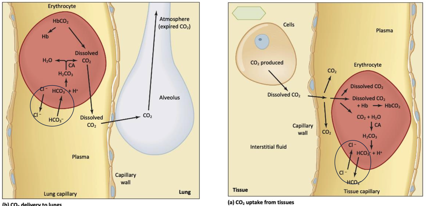
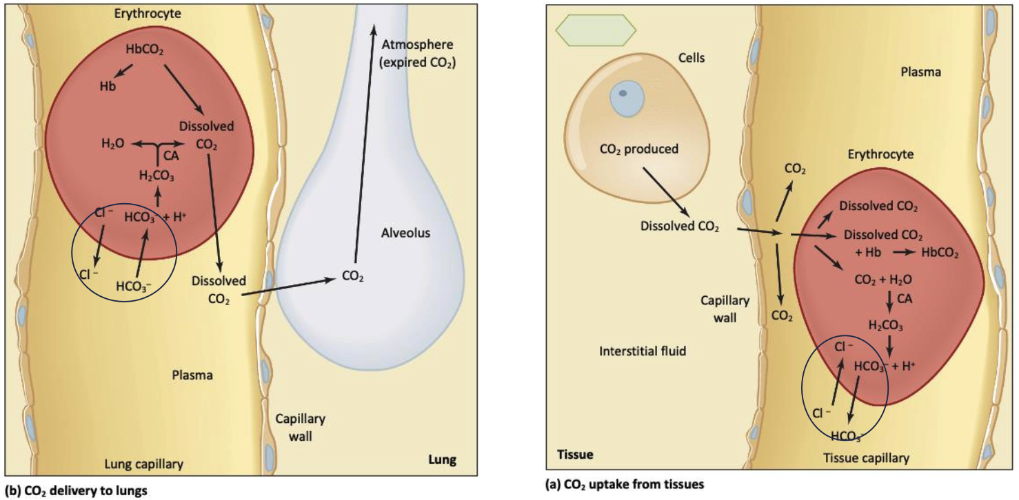
Hemoglobin can binds to oxygen, CO2
CO2 is transported out via Hb(30%), as HCO3- (60%), and dissolves in fluids (10%)
Carbonic Anhydrase (CA) facilitate CO2+H2O to HCO3
Anion Exchanger 1 (AE1) transport 1 HCO3- while Cl- in the opposite direction out/in of hemoglobin which HCO- leave Hb near cell and enters near alveoli
The Hb can be remained bind to 75% to 10% binded with oxygen
Increased metabolism (increased CO2 content and/or H+) in peripheral tissue increase O2 release by decrease Hb affinity for O2.
Negative feedback loop 1
controlled variable: peripheral H+
effectors: muscle controlling ventilation
sensor: chemoreceptor neurons
controller: brainstem, medulla onlongata
actuating signal: motor neurons
increasing ventilation increase CO2 release and O2 brought in
Negative feedback loop 2
controlled variable:central H+
effectors: muscle controlling ventilation
sensor: chemoreceptor neurons
controller: brainstem, medulla onlongata
actuating signal: motor neurons
increasing ventilation increase CO2 release and O2 brought in
Negative feedback loop 3
controlled variable: peripheral O2
effectors: muscle controlling ventilation
sensor: chemoreceptor neurons
controller: brainstem, medulla onlongata
actuating signal: motor neurons
increasing ventilation increase CO2 release and O2 brought in
ErythroPOietin, increase RBC level (long term level)
Sensor: peritubular interstitial cells in kidney
Controller: Peritubular Interstitial Cell in kidney
Actuating signal: EPO
Effector: RBC precursor cells in bone marrow
Controlled variable: PO2 level in kidney interstitial space