Ch6 Renal System
Copyright © 2025 Mark Song
Kidney
Input: renal artery
Output: renal vein and ureter
A functional unit, the nephron
the blood flow through artery, afferent arteriole, glomerular capillary, efferent arteriole, peritubular capillary, vein
the urine is generated at bowmen’s space, that is attached to glomerular capillary via glomerular filtration.
Then tubular secretion give more from peritubular capillary to tubule and tubular reabsorption some back to the capillary
the remaining go to urinary excretion
Location:
Glomerulus and Bowman’s capsule (cortex)
Proximal tububle, cortex
Descending loop of henle (cortex to medulla)
Ascending loop of henle (medulla to cortex)
Distal tubule, from Ascending loop of henle to medullary collecting duct (cortex)
Glomerular Filtration
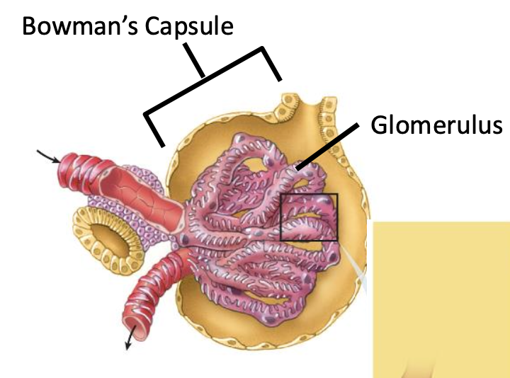
Pressure pushes water, salts, glucose, and urea out (no cell no protein)
Proximal Tubule Reabsorption
The lumen was attached to the epithelial cell (apical side) and all glucose were reabsorbed
the basolateral side of the tubular epithelial cell are to the peritubular capillary
Tubular epithelial cell
with primary transport Na/K ATPase (neither)
with Sodium GLuocse Transporter SGLT1, and SGLT2 (apical), secondary active transport (cotransport with Na conc. gradient)
with Cl- channel (apical and basolateral)
with AQ1 channel for water (apical and basolateral), osmosis
with Glu1 and Glu2 channel for glucose (basolateral), diffusion
Glucose, Cl-, and water are from tubule to the interstitial space
The units lays in the cortex (outside) and the medulla (inside) and the urine go to the ureter for urinary bladder
Descending limb of the loop of Henle
with primary transport Na/K ATPase (neither)
with AQ1 channel for water (apical and basolateral), osmosis
water are from the tubule to the interstitial space (high salt conc)
Ascending limb of the loop of Henle
with primary transport Na/K ATPase (neither)
with NKCC2 that transport Na, K, Cl (apical) all enters the cell
with K channel (apical)
with Cl- channel (basolateral)
Na, K (less than Na) flows from tubule to apical, Cl flow from tubule to interstitial space
this makes the interstitial space high salt conc for descending limb of the loop of Henle
Distal (convoluted) tubule
with primary transport Na/K ATPase (neither)
with sensitivity to hormones
with primary transport Na/K ATPase (neither)
with Epithelial Na Channel (ENaC) (apical)
with Cl- Channel (basolater and apical)
Na and Cl are flowed from the tubule to the interstitial space
Medullary collecting duct (in medulla)
with primary transport Na/K ATPase (neither)
with AQ2 channel for water (apical)
with AQ3, AQ4 channel for water (basolater)
water are from the tubule to the interstitial space (high salt conc)
Vasopressin / Antidiuretic Hormone (ADH)
it binds to Vasopressin Receptor (GPRC), activate cAMP, PKA, and exocytosis of AQ2
increase AQ2 expression on apical side membrane
increase water reabsorption
Negative Feedback Loop
Sensor: Osmoreceptor neurons in the hypothalamus, fire more if dehydrated
Controller: Neuronal network in hypothalamus
Actuating signal: ADH, Vasopressin released by posterior pituitary
Effectors: AQ2 expression level in medullary collecting duct
Controlled variable: Osmolarity of interstitial space in hypothalamus
Diabetes Insipidus
Neurogenic: loss of function mutation affecting hypothalamus
Nephrogenic: mutation affecting collecting duct cells
Macula Densa can sense salt content which is located at the end of the ascending limb of the loop of Henle and the beginning of distal tubule
Juxtaglomerular cells is the cell close to Macula Densa on the afferent arteriole
Macula Dense senses low salt content and triggers a feedback look to increase salt reabsorption
Controller and sensor, Macula Dense (releases ProstaGlandin when low tubular salt level)
Actuating signal: PG, Juxtaglomerular cells release renin, angiotensin I (cleved from angiotensiogen that was secreted from liver by renin), angiotensin II (modified by Angiotensin-converting enzyme (endothelium) from angiotensin I), adrenal cortex releases aldosterone
Effector, epithelial cells lining Distal Tubule and Cortical collecting duct
Controlled Var.: Salt content of the beginning of the Distal tubule
When aldosterone binds,
Increase transciption of Na/K ATPase and ENac